By Uruguay Clinic
When it comes to rehabilitation after an anterior cruciate ligament (ACL) injury, one of the most important aspects for restoring proper knee function is the recovery of muscle synergy between the vastus medialis (VMO) and the rectus femoris.
In this guide, you’ll learn how to identify neuromuscular imbalances after injury, which tests to use with surface electromyography (EMG), and how to personalize treatment based on the data you collect.
By the way, want to start using EMG with your ACL patients and improve your outcomes today?
👉 Contact us here to find out how.
What’s the Expected Synergy in a Healthy Knee?
During knee extension movements (e.g., squats, lunges, or isometric extensions), the most efficient neuromuscular pattern is:
✅ Vastus medialis > Rectus femoris
Stronger VMO activation is essential because this muscle helps keep the patella aligned during movement.
By pulling it slightly medially, it prevents lateral drift—critical for reducing imbalances in the extensor mechanism and minimizing the risk of femoropatellar pain or injury.
However, due to its biarticular nature, the rectus femoris tends to dominate in many patients unless synergy is actively retrained.
What Happens After an ACL Injury?
Whether post-surgical or non-operative, it’s common to observe disruptions in this muscle synergy:
- VMO inhibition (arthrogenic or central)
- Rectus femoris inhibition, or…
- Rectus femoris dominance in the absence of proper medial control
Consequences of Altered Synergy
- ❌ Joint instability
- ❌ Anterior knee pain
- ❌ Slow or stalled rehab progress
- ❌ Higher risk of reinjury or functional failure
How to Assess VMO–Rectus Femoris Synergy with EMG
To detect these alterations objectively and personalize rehab, follow this clinical protocol:
STEP 1: Measure Activation of Both Muscles
Use surface EMG to record the activation levels (in microvolts) of the VMO and rectus femoris during functional tasks.
STEP 2: Compare Healthy vs. Affected Side
Look for:
- Absolute differences in activation
- Altered synergy pattern (e.g., rectus femoris > VMO)
- Hidden deficits during real-life or sport-specific tasks
Recommended Tests for Synergy Assessment
1️⃣ Isometric Knee Extension
Goal: Identify whether the VMO is less active than the rectus femoris on the injured side.
Common Results:
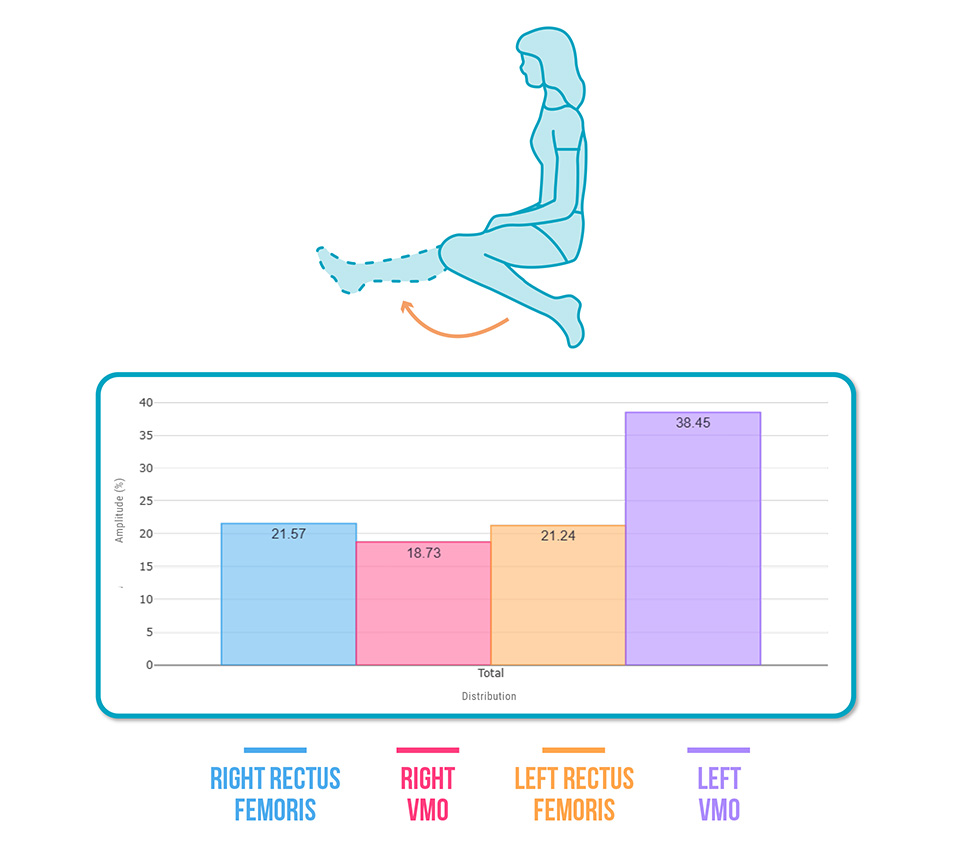
- – Right rectus femoris > Right VMO (❌)
- – Right VMO << Left VMO (❌)
Diagnosis: VMO inhibition on the affected side
2️⃣ Weighted Squat
Goal: Assess motor patterns during a functional movement.
Common Results:
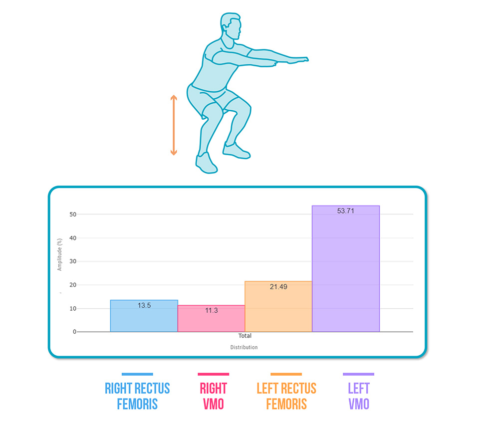
- – Right rectus femoris > Right VMO (❌)
- – Both muscles inhibited compared to healthy side (❌)
Diagnosis: Protective pattern with bilateral deficits on the injured side
3️⃣ Weighted Lunges
Goal: Analyze a more dynamic and demanding gesture.
Common Results:
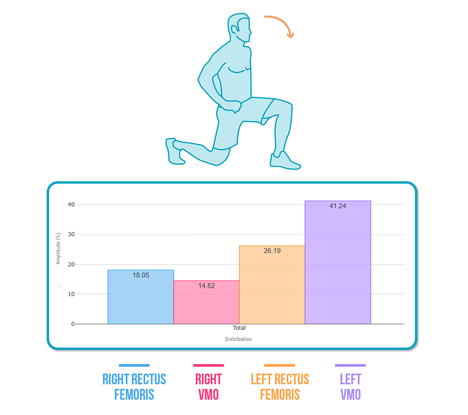
- Right rectus femoris > Right VMO (❌)
- Reduced activation of both muscles vs. healthy side (❌)
Diagnosis: Ongoing inhibition during functional load-bearing tasks
How to Intervene Based on EMG Findings
Once the deficit is identified, build your treatment plan around these pillars:
1. Strength Training
- – Targeted quadriceps exercises: isometric, isotonic, partial weight-bearing
- – Unilateral and functional work: lunges, step-ups, leg press with feedback
2. Neuromuscular Reeducation with Biofeedback
- – Real-time visualization of VMO activation
- – Correct compensations (e.g., rectus femoris dominance)
- – Teach the patient to “feel” the VMO working in each rep
3. Percutaneous Neuromodulation or E-Stim
- – Use as a complementary technique to reactivate inhibited muscles
- – Improve cortical response and muscle-brain connection
Conclusion
Assessing VMO–Rectus Femoris synergy after ACL injury is essential for effective rehabilitation.
It’s not just about regaining strength—it’s about ensuring a correct, efficient motor pattern.
EMG gives you the data you can’t see with the naked eye.
With it, you can detect subtle deficits, adjust your plan from session one, and make every rep count.
Want to learn how to integrate EMG-based evaluation into your sessions?
👉 Start using muscle assessment technology today. Book a call and learn how to integrate EMG into your clinical practice step by step.