Does your patient have persistent groin pain that just won’t go away?
You’re likely dealing with a case of pubalgia, a condition that affects many athletes and active individuals, often linked to repetitive hip flexion movements.
The problem is that you may not be evaluating what really matters.
Even when the pain is clear, its origin often isn’t. There may be hidden muscular imbalances, like poor activation of the rectus femoris or overload of the adductor longus, which cause tension and friction at the pubic area.
If not detected in time, these imbalances can perpetuate pain and hinder full recovery.
The good news: you can identify and address the issue in real time.
Thanks to surface electromyography (EMG), you can objectively evaluate how these muscles behave during hip flexion—helping you spot compensations, adjust treatment, and reduce the risk of relapse.
Want to learn how to detect these patterns and make muscle-specific decisions based on real data?
Book a free 10-minute call with an EMG expert physiotherapist. No jargon. 100% personalized.
In this post, you’ll learn how to properly assess this synergy using surface EMG—and how to use that information to design more effective treatment plans.
Why is it important to assess the synergy between the rectus femoris and adductor longus?
During hip flexion, the rectus femoris should be the main contributor—lifting the leg and generating most of the movement.
The adductor longus, meanwhile, acts as a stabilizer and synergist. In ideal conditions, this relationship looks like:
👉 Rectus femoris > Adductor longus
When this synergy is disrupted and the rectus femoris underperforms, the adductor longus takes on excessive load to compensate. Sustained overload here can lead to irritation at the common insertion point of the musculature on the pubic symphysis, increasing the risk of pubalgia.
What happens when the rectus femoris is inhibited?
Poor rectus femoris activation has several clinical consequences:
❌ The adductor longus overcompensates during hip flexion.
❌ This excessive coactivation increases traction at the pubic area.
❌ The result: pain, stiffness, and even secondary compensations in gait or sport-specific technique.
This pattern is not always visible to the eye or detectable through manual strength testing.
That’s why using objective tools like surface EMG is essential.
Clinical Case: Patient with Groin Pain
During a supine hip flexion test, we used EMG to analyze rectus femoris and adductor longus activation in both legs.
Results:
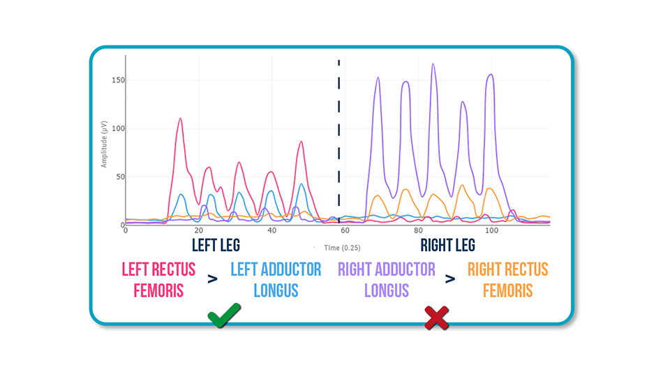
The pubalgia was linked to poor activation of the right rectus femoris and compensatory overactivity of the adductor longus.
This pattern was creating continuous overload on the pubic insertion.
What to do after detecting this pattern?
Once the altered pattern is identified, you can create a more targeted intervention plan. Here are some key strategies:
Targeted rectus femoris activation
Design hip flexion exercises that isolate and progressively activate the rectus femoris, avoiding dominant involvement of the adductor longus.
EMG biofeedback
Help the patient visualize, in real time, whether they’re activating the correct muscle. This improves motor relearning and enhances exercise effectiveness.
Technique correction in compound movements
In movements like lunges, step-ups, or running, observe whether the activation pattern remains dysfunctional.
Load control and progressive reconditioning
Temporarily reduce load on the pubic region and plan a gradual return based on symptoms and EMG activation data.
Benefits of Evaluating Muscle Synergy with EMG
- – Clinical objectivity: Identify real imbalances, even when strength appears normal.
- – Personalized treatment: Choose the right exercises and avoid those that reinforce compensation patterns.
- – Higher patient engagement: Seeing their own muscle activation increases patient commitment to therapy.
Conclusion
Pubalgia isn’t always just a structural issue—it’s often the result of inefficient muscular patterns.
By evaluating the synergy between the rectus femoris and adductor longus, you can uncover imbalances before they become chronic or complicate recovery.
If you want to improve your assessments and communicate more clearly with your patients, surface EMG lets you analyze what’s not visible and make better-informed clinical decisions.
Want to learn more about applying EMG in real cases?
👉 Book a free 10-minute call with a physiotherapist specialized in EMG. No jargon. 100% personalized.
See you in the next post 🙂