Do you know why your patient has reduced plantar flexion mobility? Or why they lack strength? The answer may lie in the muscles contributing to that movement. It is very likely that some of the muscles that should be leading it are not doing so.
When the plantar flexion synergy is disrupted, the system redistributes the work among muscles that are not designed to take on that role. The result is a patient who trains, who puts in effort, but whose motor pattern continues to organise itself around compensation rather than around the primary muscle.
And that does not improve with more load. It improves when you identify which muscle is failing and correct it with data.
In this article you will discover what the expected muscle synergy is in a functional plantar flexion, how that pattern is disrupted in a real case of ankle instability and what specific changes EMG biofeedback retraining produces in the activation of each muscle.
The muscles involved in plantar flexion
Before assessing whether the pattern is correct, you need to know what the reference pattern looks like. These are the primary muscles that activate during plantar flexion and the role each one plays:
1. Soleus. It is the primary mover of the movement, especially under load. Its deep position and predominantly slow-twitch fibre composition make it particularly relevant in endurance activities such as walking, running or postural maintenance.
2. Medial and lateral gastrocnemius. They activate more when the knee is in extension, because the gastrocnemius crosses two joints. With the knee extended they are powerful plantar flexors; with the knee flexed, their contribution decreases and the soleus takes on a greater role.
3. Peroneals (peroneus brevis and longus). They provide lateral stability to the ankle and control the relationship between inversion and eversion during movement. In functional plantar flexion they work as active stabilisers of the lateral longitudinal arch.
4. Tibialis anterior. Under normal conditions it acts as an antagonist to plantar flexion. Its presence as an activator during this movement is a warning sign: it may be functioning as a compensatory stabiliser when the primary pattern is disrupted.
The expected synergy in functional plantar flexion
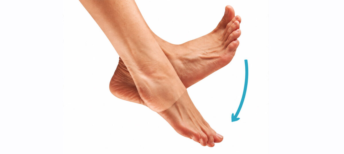
In a functional plantar flexion, the activation order and relative level of each muscle should follow this hierarchy:
Soleus > Gastrocnemii > Peroneals > Tibialis anterior
This synergy varies slightly depending on the type of load, the speed of movement and the type of weight-bearing in the exercise.
With eccentric load, the soleus and gastrocnemii increase their demand. With single-leg support, the peroneals become more prominent. But in all cases, the soleus should lead the movement and the tibialis anterior should remain in a secondary, stabilising role.
When that hierarchy is reversed or distorted, you are looking at a disrupted pattern that EMG can detect before it generates symptoms, or, more often, that explains why the symptoms already present are not resolving with standard treatment.
Real case: patient with ankle instability
This is a real example of what EMG reveals when the plantar flexion pattern is disrupted in a patient with plantar fasciitis.
The findings before the intervention showed two main alterations:
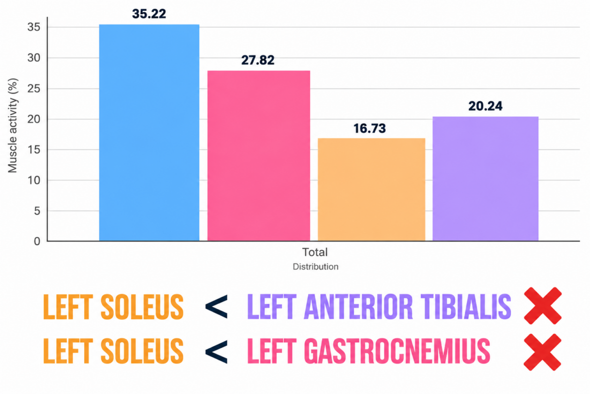
The soleus has an activation deficit that is contributing to a reduced capacity to generate force in plantar flexion.
Retraining the pattern with EMG biofeedback
After the EMG biofeedback intervention, the activation pattern reorganised towards the expected functional synergy:
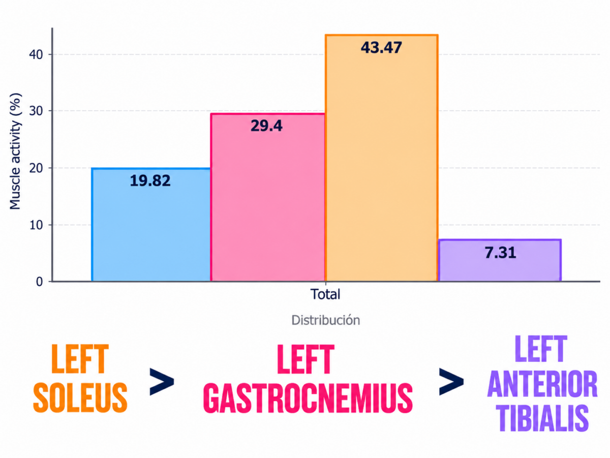
The soleus doubled its activation level. The tibialis anterior reduced its contribution to less than a third of its initial value.
In a single biofeedback session, the compensatory pattern was reversed in a measurable way.
This is what EMG biofeedback achieves that no verbal instruction can match with the same efficiency: the patient sees in real time which muscle they are activating and can modulate their own motor pattern based on that information. Motor learning is faster because the feedback is immediate, specific and objective.
Frequently asked questions
Does the plantar flexion synergy change with knee position? Yes. With the knee in extension, the gastrocnemii contribute more because they are at an optimal length for force generation. With the knee flexed, the soleus takes on a greater role because the gastrocnemii are in a shortened position. This principle is important for designing the assessment protocol: evaluating plantar flexion in both positions gives you a more complete picture of each muscle’s function.
Should the tibialis anterior always be inhibited during plantar flexion? Not completely. A low level of tibialis anterior activation is normal as stabilising coactivation. The problem arises when it exceeds soleus activation, which is exactly what happens in the clinical case described. A tibialis anterior > soleus ratio during plantar flexion is a clear sign of a compensatory pattern.
How many biofeedback sessions are needed to change the pattern? It depends on the degree of dysfunction and the patient’s motor learning capacity. In cases like the one described, a single session can produce measurable changes. However, for those changes to consolidate into an automated motor pattern, several sessions are generally needed with progressive practice under conditions of greater load and complexity.
Can this protocol be used for other conditions such as Achilles tendinopathy? Yes. Achilles tendinopathy can also have a similar synergistic dysfunction component, particularly when there is gastrocnemius dominance over the soleus. Assessing the plantar flexion synergy with EMG is equally informative in that context and can guide the eccentric loading programme towards the muscles with the greatest activation deficit.
Conclusion
Plantar flexion is neither a simple nor a uniform movement. It has an expected muscle synergy that, when disrupted, explains many of the foot and ankle conditions that do not improve with standard treatments.
The ankle instability case is a clear example: an inhibited soleus and an overactive tibialis anterior are findings you can only detect by measuring. And once you detect them, EMG biofeedback gives you the tool to correct them in an objective and verifiable way.