By Alex Ballester
After knee surgery, one of the most common rehabilitation challenges is arthrogenic muscle inhibition (AMI) in the quadriceps. This dysfunction prevents proper muscle activation, delaying recovery and affecting the patient’s functional performance.
How Does AMI Affect Recovery?
- ⚠️ Persistent pain that makes muscle activation difficult.
- ⚠️ Muscle atrophy and altered neuromuscular patterns, affecting motor control.
- ⚠️ Delayed strengthening, prolonging the rehabilitation process.
Practical Example: Why You Should Measure Muscle Function in ACL Recovery
Imagine a patient recovering from anterior cruciate ligament (ACL) surgery. At first glance, their knee seems to be healing well, but despite strengthening efforts, their quadriceps isn’t responding as expected. The patient experiences early fatigue and knee instability when descending stairs.
If you don’t measure muscle function, it’s easy to assume they just need more strength training. However, the real issue might be AMI, where the vastus medialis fails to activate properly due to post-surgical inflammation and neuromuscular changes.
If this dysfunction goes undetected, the patient may develop compensations, delay their recovery, and increase the risk of re-injury.
If you want to achieve outstanding success in overcoming AMI, applying advanced strategies to re-educate activation patterns and optimize quadriceps function, you’re in the right place. Keep reading.
Neuromuscular Response: What’s Failing in This Knee Extension?
By the way, discover how mDurance EMG can help you improve your results.
Watch this knee extension exercise and answer: Which muscle appears most active?
- a) Vastus Medialis
- b) Vastus Lateralis
- c) TFL

Observing the knee extension exercise on a treatment table, we detect that:
➡️ Tensor Fasciae Latae (TFL) is the most active muscle, surpassing Vastus Lateralis, Vastus Medialis, and Rectus Femoris.
Neuromuscular Pattern Error:
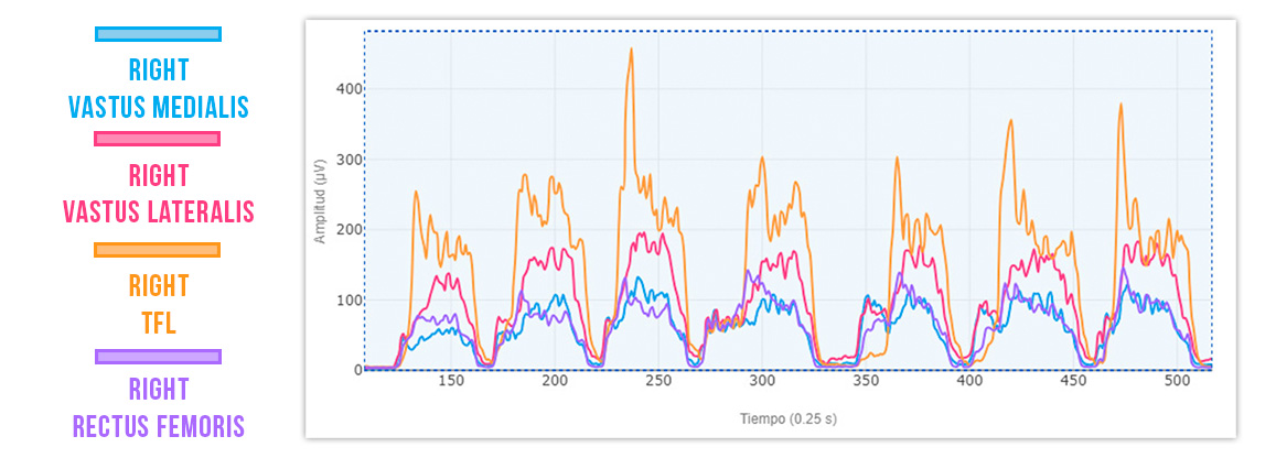
- ❌ TFL > Vastus Lateralis, Vastus Medialis, and Rectus Femoris
- ❌ TFL is overactive, despite playing a secondary role in this exercise.
- ❌ Vastus Medialis inhibition, compromising knee stability and extension efficiency.
This incorrect pattern prevents effective recovery from AMI, as the quadriceps isn’t functioning optimally.
Re-Education with EMG Biofeedback
Surface Electromyography (EMG) is the ideal tool for correcting altered neuromuscular patterns through real-time biofeedback. With this technology, your patient can visualize their muscle activation and consciously adjust their effort, achieving more efficient movement execution.

Thanks to biofeedback, in just a few minutes, the patient can re-educate their activation pattern:
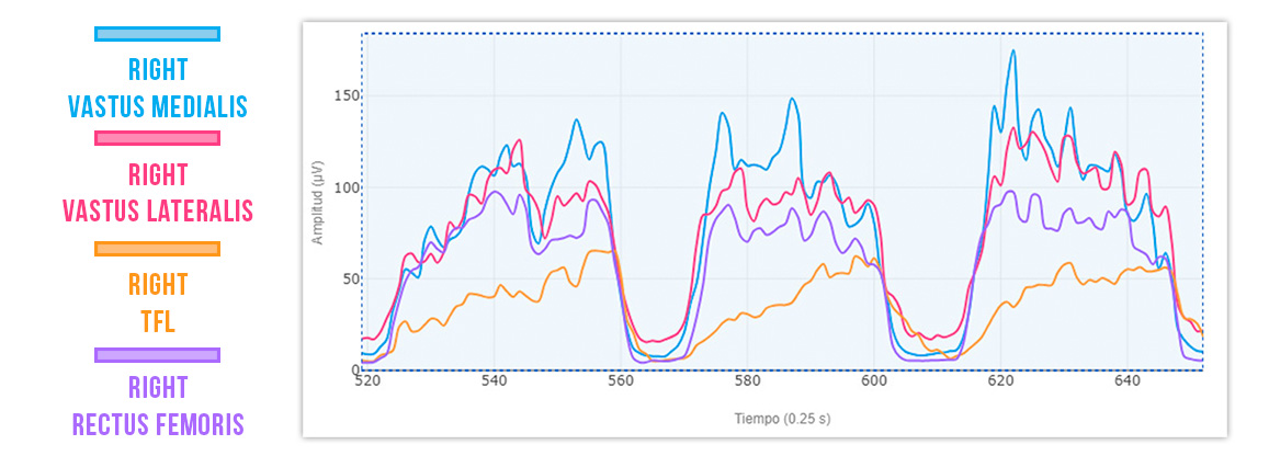
- ✅ Vastus Medialis > Vastus Lateralis and Rectus Femoris
- ✅ Rectus Femoris > TFL
- ✅ Greater efficiency in knee extension
Now, compare post-biofeedback Vastus Medialis activation with the initial graph:
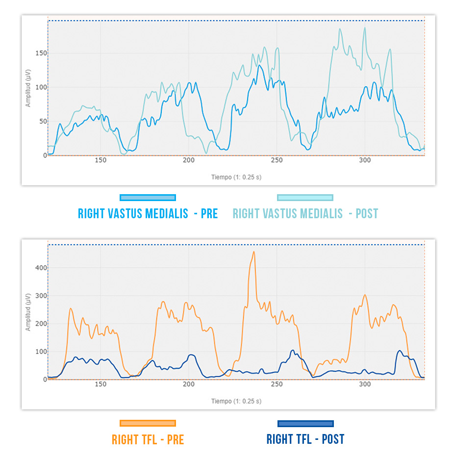
- ✔️ Increased Vastus Medialis activation.
- ✔️ Significant reduction in TFL involvement.
This process optimizes AMI recovery, ensuring proper muscle activation and reducing unnecessary compensations.
Conclusion
Arthrogenic muscle inhibition (AMI) after ACL injury or surgery can delay recovery if not detected and corrected in time. Neuromuscular re-education is key to restoring proper activation patterns and avoiding compensations that impact knee function.
EMG Biofeedback allows you to:
✔️ Prevent incorrect muscle activations in different exercises.
✔️ Customize exercises based on each patient’s needs.
✔️ Maximize execution efficiency, accelerating functional recovery.
Discover how mDurance EMG can help you improve your results.
See you in the next post 🙂