Do you know whether your patient correctly dissociates the pelvis from the spine when moving?
If you do not have an objective way to measure it, you are probably starting lumbar rehabilitation without knowing exactly where your patient’s real baseline is.
Many patients with chronic low back pain or pelvic instability report pain when moving or exercising, activate the lumbar region more than the gluteus during hip extensions and have a lack of pelvis-spine dissociation that perpetuates the problem.
To the naked eye, the movement may look acceptable. But underneath there is an altered neuromuscular pattern that no clinical observation can quantify with precision.
In this article you will discover the mDurance Lumbopelvic Dissociation Protocol: five exercises with which you can detect in real time which muscle is leading the movement, where compensations appear and how your patient’s lumbopelvic control is organised before starting rehabilitation.
Why lumbopelvic control is the first variable you should measure
When dissociation between the pelvis and spine is lost, the consequences are not limited to low back pain. The entire system reorganises itself around that dysfunction:
❌ Greater joint stress in the lumbar segments, which translates into recurring pain that is difficult to resolve as long as the motor pattern does not change.
❌ Gluteal activation deficit, because when the spinal erector dominates the movement, the gluteus maximus is pushed into the background. The result is reduced capacity to generate hip force and a posterior chain that does not function as it should.
❌ Compensations in other muscles, which increase the risk of injuries in the knee, hip or even the thoracic spine in the medium term.
The problem is that these patterns are not diagnosed by observing. They are diagnosed by measuring. And the time to do it is before prescribing the first exercise, not after the patient has spent weeks without improving.
The mDurance Lumbopelvic Dissociation Protocol
The protocol is designed to detect three types of findings in real time:
🎯 Which muscle is leading the movement: gluteus maximus or spinal erector?
🎯 Whether there is excess activation or lumbar stiffness during the movement.
🎯 Where losses of control or lumbopelvic compensations appear.
It consists of five exercises, each with a specific clinical goal and a set of findings that EMG can detect and that observation cannot quantify.
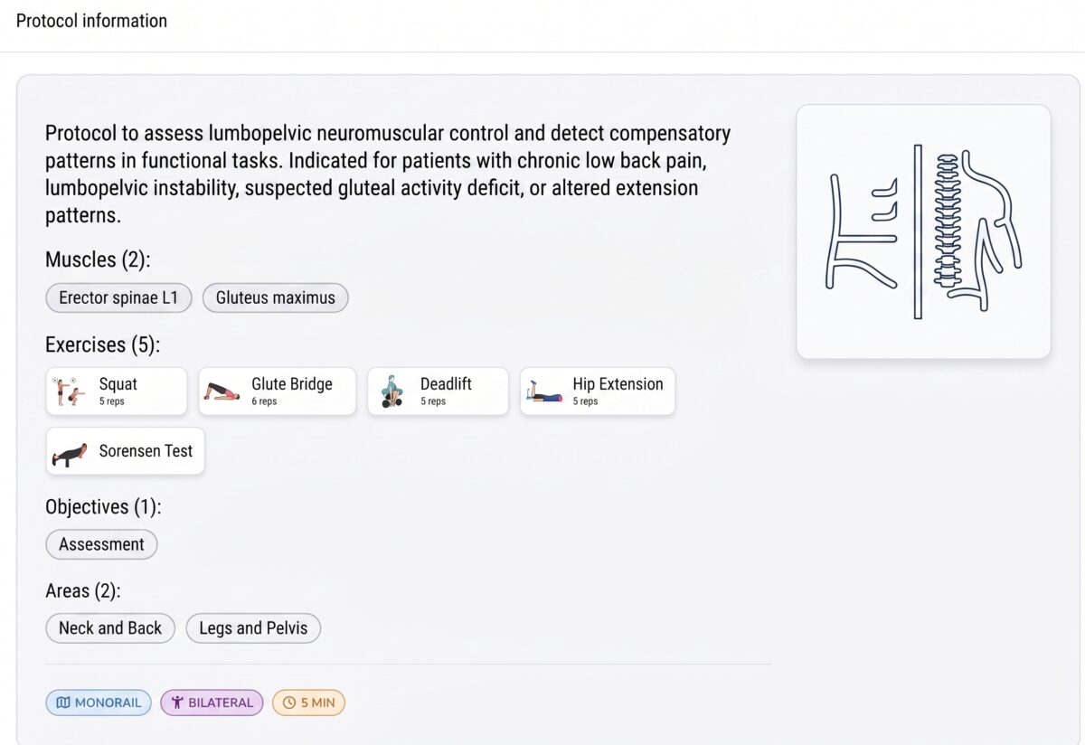
Exercise 1: Squat (8 repetitions)
Goal: assess lumbopelvic control during a global flexion-extension movement.
The squat is one of the most informative tests for lumbopelvic control because it involves both the spine and the hip throughout their full range of motion. EMG allows you to see the activation sequence in real time and detect two especially common patterns:
Excessive spinal erector activation at the start of the descent. When the erector dominates the eccentric phase, it indicates that the spine is controlling the descent rather than the hip. The correct pattern requires the gluteus and hamstrings to lead the lowering phase with the spine in a neutral position.
Low gluteus participation when rising to standing. If the spinal erector continues to dominate the ascent, the gluteus maximus is not generating the hip extension it should. This finding is especially relevant in patients who report that the squat loads their lower back.
Exercise 2: Glute bridge (8 repetitions)
Goal: analyse the activation sequence during hip extension in an unloaded position.
The glute bridge is the most direct test for assessing whether the gluteus maximus assumes its primary function in hip extension or whether the lumbar muscles compensate for it. The most common findings are:
Gluteus that activates late or with low intensity. EMG shows the activation timing of each muscle with precision. A gluteus that responds with a delay or that does not reach sufficient activation levels is a clinical finding that no manual test can provide with the same precision.
Lumbar muscles compensating from the start and throughout the movement. When the spinal erector activates before or at the same time as the gluteus in the glute bridge, the motor pattern is inverted. The spine is doing the work that the hip should be doing.
Difficulty maintaining a neutral pelvis during extension. EMG can detect the micro-adjustments the muscular system makes to hold the position, revealing when pelvic stability depends on lumbar tension rather than active gluteal control.
Exercise 3: Deadlift (8 repetitions)
Goal: observe eccentric control and return from trunk flexion.
The deadlift assesses a pattern that the squat cannot evaluate in the same way: control of the hip hinge under load. It is especially useful for detecting alterations in the timing between the pelvis and spine during the lowering and rising movement.
Overactivated spinal erector during the ascent. If the erector activates excessively during the concentric phase of the deadlift, extension is occurring primarily in the spine and not in the hip. This is the most frequent mechanical origin of low back pain associated with the deadlift.
Lack of gluteus-erector coordination on the way up. The ideal pattern requires the gluteus and erector to work in a coordinated way during extension. When the erector activates significantly before the gluteus, the lumbar spine is absorbing the load that the hip should be managing.
Altered timing between pelvis and spine. EMG detects whether the pelvis and spine are moving in a discoordinated way, which is one of the most sensitive markers of lumbopelvic dysfunction.
Exercise 4: Hip extension (8 repetitions)
Goal: detect whether the gluteus maximus is performing its primary function or whether the lumbar muscles are compensating.
Hip extension in four-point kneeling or prone is the purest test for assessing lumbar vs gluteal dominance in extension. Without the variable of knee movement or body weight, EMG can isolate with greater precision which muscle is leading the extension.
Spinal erectors dominating the movement. In many patients with chronic low back pain, hip extension primarily activates the lumbar muscles rather than the gluteus. This pattern is common and difficult to detect without EMG.
Gluteus with deficit and/or delayed activation. A gluteus that is late to hip extension is an inhibited gluteus. EMG shows that delay with a precision that no manual clinical test can match.
Difficulty maintaining control at the end of the range. Loss of lumbopelvic stability at the end of hip extension indicates that the muscular system does not have sufficient capacity to control that range. It is one of the most frequent and least assessed deficits in patients with low back pain.
Exercise 5: Sorensen test (maximum time)
Goal: assess spinal erector endurance under sustained load conditions.
The Sorensen test is the reference assessment for evaluating the endurance of the lumbar extensor musculature. Unlike the previous exercises, it does not assess the activation pattern during movement but rather the capacity to sustain activation over time.
Early erector fatigue. If the erector fails earlier than expected for the patient’s age and profile, it indicates insufficient lumbar endurance capacity for the demands of their daily or sporting life.
Asymmetry between erectors. EMG allows comparison of the activation of the right and left spinal erector during the test. A significant asymmetry is a clinically relevant finding that can explain unilateral pain patterns or load compensations in the spine.
Frequently asked questions
Is this protocol suitable for patients in the acute phase? Not in all cases. The glute bridge and prone hip extension may be well tolerated even in subacute phases, but the squat, deadlift and Sorensen test require the patient to have sufficient load capacity. The protocol is designed primarily for the assessment phase prior to active rehabilitation or at the start of reconditioning.
How many EMG channels do I need for this protocol? With four channels you can assess the gluteus maximus and spinal erector simultaneously, which are the two primary muscles in the protocol. If you want to add the gluteus medius or hamstrings, you would need more channels. mDurance works with four EMG channels.
Can I use this protocol to monitor treatment progress? Yes, and this is one of its most valuable uses. Repeating the protocol at different points in the process allows you to objectively demonstrate whether the lumbopelvic pattern is changing, whether the gluteus is gaining prominence over the erector and whether compensations are decreasing. It is a way of demonstrating progress with data, not just with the patient’s perception.
What do I do if I find that the erector dominates in all the exercises? This is one of the most common findings and the clearest starting point for designing the programme. It means you need to re-educate gluteus maximus activation before progressing in load. You can work with EMG biofeedback to help your patient gain strength while avoiding compensations.
Conclusion
Lumbopelvic control is the foundation of any lumbar rehabilitation programme that aims to be effective in the medium term. Without knowing whether the patient dissociates the pelvis from the spine, whether the gluteus maximus leads hip extension or whether the erector is compensating in all movements, you are prescribing exercises without knowing exactly what is failing.
The mDurance Lumbopelvic Dissociation Protocol gives you that information in 15 minutes, with objective data in real time. Five exercises, two key muscles and a clear picture of your patient’s neuromuscular pattern before starting treatment.
Because the difference between a programme that works and one that does not is often not in the exercises. It is in knowing what is failing before choosing them.