Do your patients perform horizontal rows without truly activating the right muscles?
At first glance, the movement might look fine. But there’s a critical difference between a good pattern and one that leads to overload—and it’s invisible without electromyography.
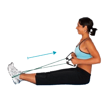
The horizontal row with a resistance band is one of the most common exercises for targeting posterior trunk muscles, improving posture, and treating shoulder and neck pain. However, its effectiveness depends on proper technique and, more importantly, a balanced muscle activation pattern.
With small adjustments in technique and accurate evaluation of which muscles are actually working, this exercise can become a key tool to enhance scapular stability, reduce shoulder pain, and improve postural function.
In this guide, you’ll learn how to assess the most relevant muscle synergies during a horizontal resistance band row—using real clinical examples and EMG data—so you can apply it right away in your practice.
The result?
More objective assessments, better clinical decisions, and more effective treatments from the very first session.
Key Muscles in a Horizontal Row
When performing a horizontal row—especially with a resistance band—these are the main muscles involved:
- Latissimus dorsi: The primary mover. Responsible for generating pulling force.
- Posterior deltoid: Assists in the final phase of the row and helps stabilize the shoulder.
- Lower trapezius: Essential for scapular retraction and depression. Key to scapular stability.
- Upper trapezius: Has a stabilizing role but should be controlled. Overactivation can lead to unwanted neck and shoulder tension.
Expected Synergy
The ideal activation sequence in a horizontal resistance band row should follow this pattern:
Latissimus dorsi > Posterior deltoid > Lower trapezius > Upper trapezius
This order reflects efficient force distribution:
The lats lead the pull, the posterior deltoid assists without dominating, and the traps activate in a balanced way to stabilize the movement.
Clinical Case: Patient with Shoulder Pain
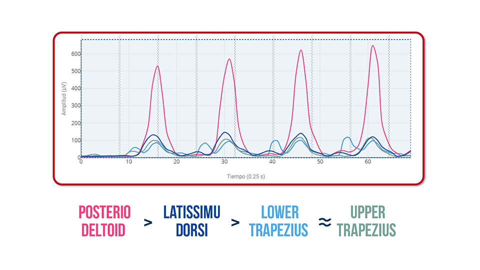
Observed Pattern:
Posterior deltoid > Latissimus dorsi > Lower trapezius = Upper trapezius ❌
Findings:
- – Overactive posterior deltoid → Takes on more load than necessary, potentially leading to fatigue or joint overload.
- – Underactive latissimus dorsi → The primary mover isn’t doing its job.
- – Underactive trapezius → Poor scapular stabilization compromises movement efficiency.
- – Moderate upper trapezius activity → The only muscle within expected range.
This pattern may result in ongoing compensation by the posterior deltoid, increased glenohumeral joint stress, and persistent pain.
Normal Activation Pattern in a Resistance Band Row
Normal Activation Pattern in a Resistance Band Row
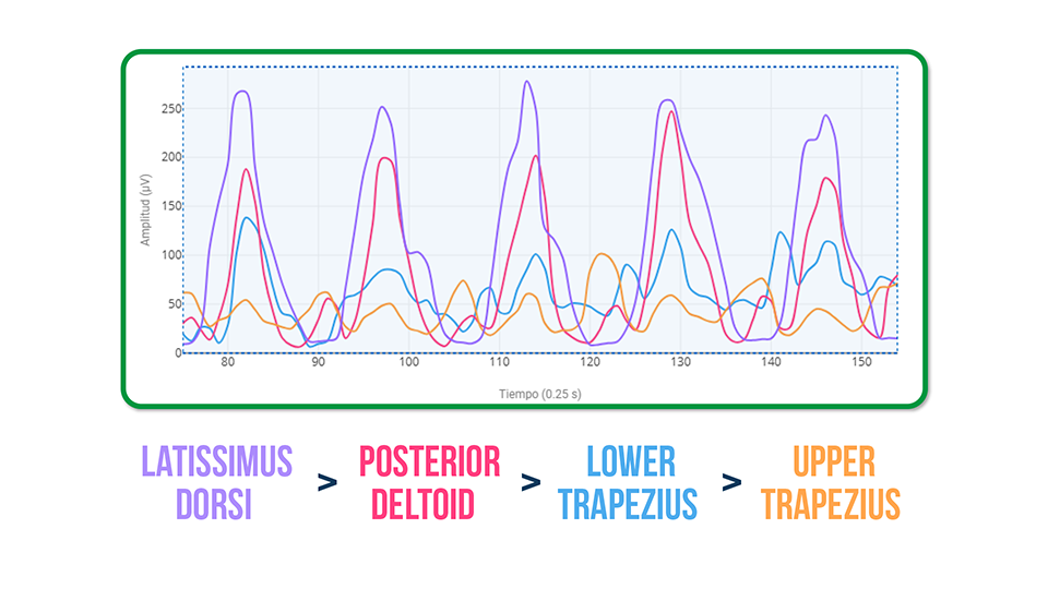
In a compensation-free execution, we see:
Latissimus dorsi > Posterior deltoid > Lower trapezius > Upper trapezius
Here, the pull originates from the lat, is distributed appropriately to the stabilizers, and the posterior deltoid doesn’t become overloaded.
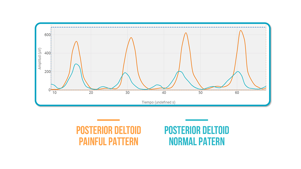
Patient with Pain vs. Normal Pattern: EMG Comparison
When analyzed using EMG:
- – The painful shoulder’s posterior deltoid shows 50% more activation than in the normal pattern.
- – This excess may lead to early fatigue and reduce the effectiveness of the exercise.
- – In contrast, the normal pattern distributes effort more effectively, with greater lat engagement and better scapular stability.
How to Use This Information
With this guide, you can:
- – Accurately assess whether the movement pattern is efficient or masked by hidden compensations.
- – Objectively measure the activation of key muscles.
- – Personalize re-education exercises to restore proper muscle synergy.
- – Use EMG biofeedback to visually and interactively correct patterns with your patients.
Conclusion
The horizontal band row is a great exercise… if done correctly.
Assessing muscle synergy between the latissimus dorsi, posterior deltoid, and trapezius muscles is essential for improving performance and reducing shoulder pain.
With tools like surface electromyography, you can spot and correct issues that would otherwise go unnoticed.
Want to learn how to use EMG in your shoulder assessments?
👉 Click here to get all the information you need to integrate electromyography into your clinical practice with mDurance