Does your patient have knee or hip pain and you are unsure whether the problem lies in how they load the muscle when climbing stairs, a ramp or uneven terrain?
The muscular pattern on an incline is completely different from that of walking on flat ground, and that difference is not always visible, measuring it will prove useful.
Many patients with patellofemoral pain, biceps femoris tendinopathy or gluteus maximus overload report that the pain appears or worsens when walking uphill, not on flat ground.
The reason may lie in how the muscles contribute: walking at a 9° incline multiplies biceps femoris activation by more than six and gluteus maximus activation by more than three compared to flat ground. If you are not assessing incline gait, you are only seeing part of the problem.
In this article you will discover which muscles increase their activation the most when walking uphill, by how much and how you can use that information to understand why your patient’s pain appears on slopes and not on flat ground.
The data: increase in muscle activation when walking at a 9° incline
The values below represent the percentage increase in activation of each muscle when walking uphill compared to flat ground, measured with EMG at a 9° incline:
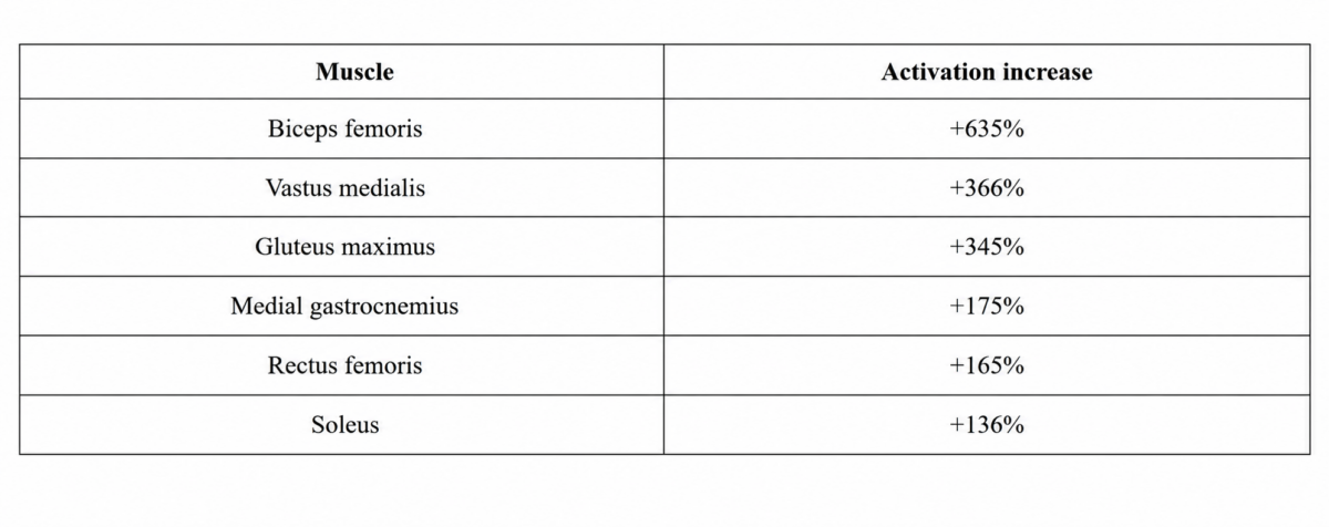
The most striking increase is that of the biceps femoris: more than six times its activation on flat ground. And the most clinically relevant point is that the three muscles with the greatest increase are hip and knee extensors, not the plantar flexors that many professionals intuitively associate with uphill effort.
Why the biceps femoris leads the ranking (and what it means clinically)
Walking uphill demands a more pronounced hip extension with each step. The biceps femoris, as a hamstring muscle, acts as the primary hip extensor in that context.
When the gradient increases, the demand on that function increases too, and the biceps femoris responds with an activation increase that has no parallel in the rest of the leg muscles.
This directly explains one of the most common patterns seen in clinical practice: the patient with posterior thigh pain who tolerates flat walking well but worsens as soon as there is a gradient. The slope demands six times more than flat ground, and if there is a tendinopathy or incomplete recovery from a previous injury, that increase brings them to the pain threshold.
How activation changes across the assessed muscles
The six muscles analysed can be grouped into two response levels:
High level (>300% increase): biceps femoris, vastus medialis and gluteus maximus. Their shared function is hip and knee extension against gravity. When the terrain tilts, their demand multiplies.
Moderate level (130-175% increase): medial gastrocnemius, rectus femoris and soleus. These also increase their activation, but to a significantly lesser extent. They remain necessary, but they are not what determines the differential effort between flat ground and a slope.
The extra effort of walking uphill does not distribute evenly across all leg muscles. It concentrates in the hip extensors.
What changes (and what does not) with speed
One of the most relevant findings in studies on incline gait is that this muscle distribution pattern is maintained even when speed changes. The relative increase between muscles does not vary significantly whether the patient walks faster or slower on the same incline.
This means that controlling speed is not sufficient to modulate the demand on the biceps femoris or the gluteus maximus. If you want to reduce the load on those muscles, you need to reduce the gradient, not just the speed.
How to use this data to understand your patient’s pain
Posterior thigh pain that appears on slopes. The 635% increase in the biceps femoris may be the explanation. If the patient has a proximal biceps femoris tendinopathy or incomplete recovery from a previous injury, the slope brings them directly to the pain threshold even when they are asymptomatic on flat ground. EMG during incline gait allows you to quantify how much activation they are generating and whether that load is compatible with their current recovery phase.
Patellofemoral or knee pain that worsens when climbing stairs or slopes. The vastus medialis increases by 366% on an incline. In patients with patellofemoral dysfunction, where the balance between vastus medialis and vastus lateralis is already compromised, that additional demand may be what reproduces the pain. Measuring the activation of both vasti during incline gait gives you direct information about whether there is asymmetry in the load on the patella.
Gluteus maximus overload with no apparent cause. A 345% increase in the gluteus maximus explains why patients who walk a lot in the city, go hiking or work on uneven terrain develop overload in that region without having changed their activity volume: they have changed the terrain. The slope multiplies the demand on the gluteus without the patient perceiving it as a qualitatively different effort.
Frequently asked questions
Does the same pattern apply to downhill walking? No. On the descent, the muscular pattern changes significantly. Eccentric quadriceps work and the demand on the gastrocnemius increase, while the gluteus maximus and biceps femoris play a different role. EMG analysis of downhill gait would produce a completely different ranking, with its own clinical implications for conditions such as chondromalacia patellae or Achilles tendinopathies.
At what gradient do these increases occur? The data correspond to a 9° incline. The magnitude of the increase varies with the angle: the steeper the gradient, the greater the demand on the hip extensors. The 9° data are a useful reference point, but the real muscular response of your patient on the gradient you are prescribing can only be obtained by measuring it.
Can incline walking be used to work the vastus medialis? Yes. The 366% increase in the vastus medialis is one of the most relevant findings in the ranking for knee rehabilitation. In patients with patellofemoral dysfunction or recovering from ACL injury, incline walking can be an interesting functional stimulus for the vastus medialis. As always, however, the indication must be adjusted to individual tolerance and verified with real activation data.
Are these data valid for all patient profiles? The general pattern (hip extensor dominance on slopes) is consistent in the literature. However, the exact magnitude of the increases varies according to age, fitness level, stride length and gait technique. EMG allows you to start from the reference values of the study and adjust the interpretation to the individual characteristics of each patient.
Conclusion
When a patient tells you that their knee or hip hurts when walking uphill but not on flat ground, remember that the slope multiplies the demand on specific muscles in a way that flat walking never reaches.
And if those muscles have a tendinopathy, incomplete recovery or an activation imbalance, the slope brings them to the pain threshold in a predictable way.
The EMG data explain it: biceps femoris ×6, vastus medialis ×3.6, gluteus maximus ×3.4 compared to flat ground. These are not performance numbers. They are the answer to why your patient’s pain appears where it appears and when it appears.