Gait is one of the most frequently repeated movements of the day, yet it’s often one of the most overlooked when it comes to evaluating muscle activation patterns.
Behind an altered gait may lie compensations, misalignments, and inefficient muscle activations that compromise function and increase the risk of injury—especially in the knee and hip.
In this guide, you’ll find a detailed explanation of which thigh muscles are most active during walking, what the ideal synergy should look like, what happens when it’s disrupted, and how to detect these issues objectively using surface electromyography (EMG).
If you work with patients dealing with knee pain, recurring injuries, or gait dysfunction, this information will change how you assess and treat them.
Key Thigh Muscles Activated During Gait
Throughout the gait cycle, several thigh muscles activate at key moments to ensure stable and efficient movement:
- Semitendinosus: Activates at the end of the swing phase to decelerate the leg and prepare for contact, aiding hip extension and knee flexion.
- Biceps femoris: Supports the semitendinosus in stabilizing the knee at the end of the swing phase.
- Vastus lateralis: Active at the end of the stance phase, helping control knee extension and slow the thigh.
- Vastus medialis: Works alongside the vastus lateralis to maintain knee alignment and stability during stance.
- Rectus femoris: Mainly active during toe-off, contributing to knee extension and propelling the leg forward.
The Expected Muscle Synergy During Gait
In a functional gait pattern, the ideal neuromuscular sequence should be:
Semitendinosus > Biceps femoris > Vastus lateralis > Vastus medialis > Rectus femoris
This pattern ensures that the hamstrings lead the control phase at the end of swing and during initial contact, while the quadriceps stabilize during stance and assist propulsion at the end.
When this synergy is altered, movement becomes inefficient and overload builds up, eventually leading to injury.
Risks of Quadriceps Dominance
When the quadriceps dominate over the hamstrings, joint balance and gait safety are compromised. This pattern can result in:
- ⚠️ Knee instability, especially at initial contact
- ⚠️ Patellofemoral overload, increasing stress on the knee joint
- ⚠️ Higher ACL injury risk, since the hamstrings help protect and stabilize the tibia during stance
Which Patients Commonly Show This Imbalance?
Quadriceps dominance is common in several clinical profiles
- Patients with chronic or recurring knee pain
- Individuals with previous hamstring injuries
- Sedentary individuals, where the hamstrings are often inhibited
- Post-surgical rehab cases (ACL repair, joint replacement, meniscectomy)
- Athletes, where muscle fatigue leads to compensation patterns
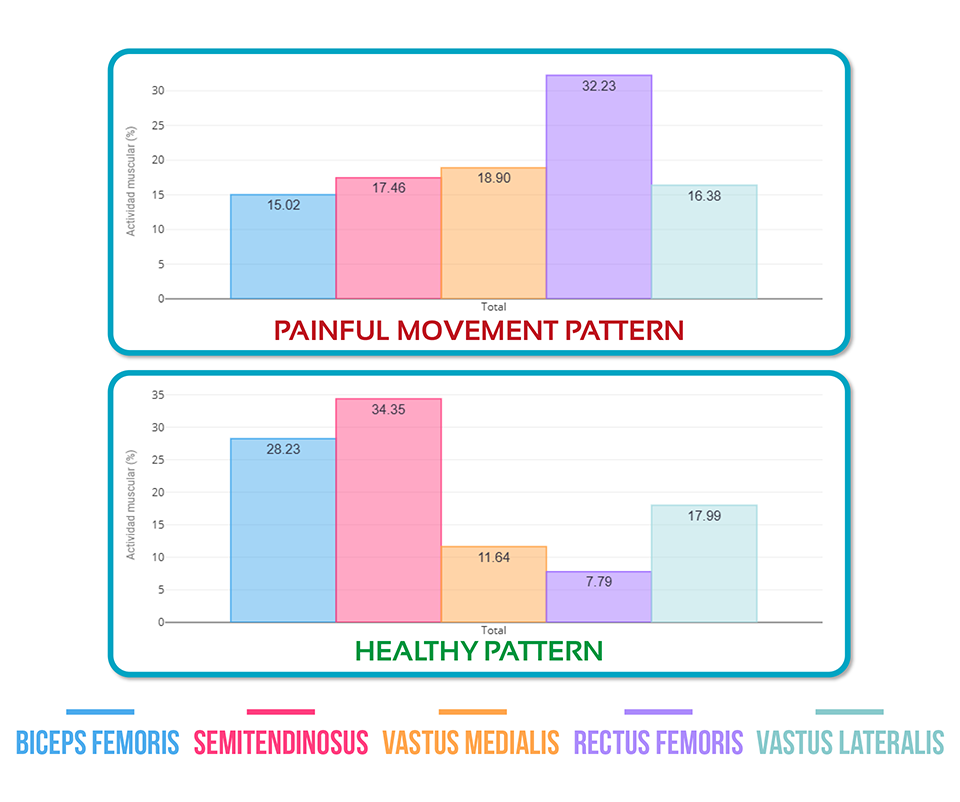
Clinical Case: Patient With Knee Pain
During an EMG assessment, the following activation pattern was recorded:
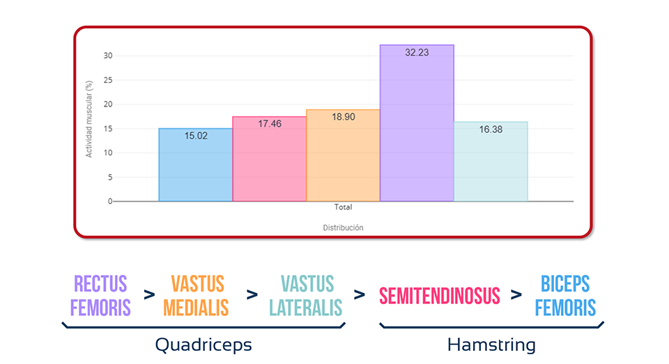
femoris ❌
This sequence shows clear quadriceps dominance and insufficient hamstring activation, which must be corrected to improve motor control.
Comparison With a Normal Gait Pattern
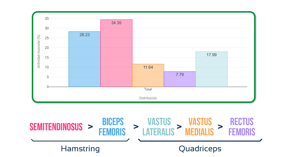
Key differences:
- Hamstrings in the ideal pattern activate twice as much as in the pain case
- The rectus femoris in the pain case activates five times more than in the healthy pattern
Conclusion: Do You Have Patients With Gait Alterations?
Ask yourself these three key questions:
- Do you have patients with visible or subtle gait abnormalities?
- Do you know which muscles are not functioning properly during walking?
- Is this affecting your treatment outcomes?
If you answered yes to any of these, it’s time to integrate tools like EMG to detect and correct these imbalances.