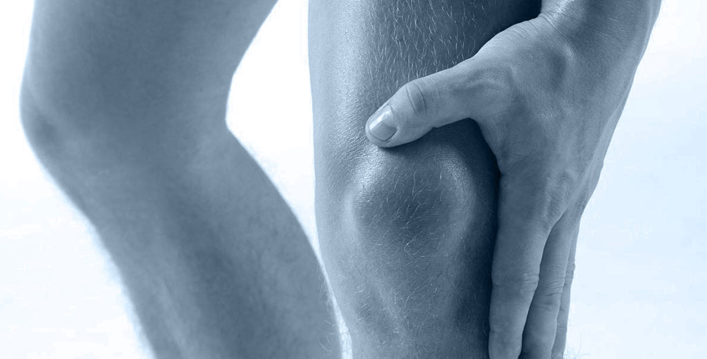
How many times have you had a patient with foot pain, gait alterations or claw toes without knowing exactly which muscle is failing?
The intrinsic musculature of the foot is one of the most difficult regions to assess objectively with standard resources. Surface electromyography (EMG) changes that.
With EMG you can measure the real activation of the foot muscles, compare sides, detect deficits and adjust treatment with data.
In this article you will learn about the 5 foot muscles you can assess, their primary function, when it makes sense to include them in your evaluation and which pathologies are commonly associated with their dysfunction.
Why assess the intrinsic muscles of the foot
The primary function of the intrinsic foot muscles is to maintain the arches and stability during gait. When one of them fails, the compensatory system redistributes the load onto other structures: other muscles, tendons, plantar fascia or joints.
The problem is that those compensations do not always generate immediate symptoms in the failing muscle. Pain appears where the compensating structure can no longer cope.
This is why directly assessing the activation of these muscles with EMG allows you to identify the origin of the problem before the compensation becomes an injury.
1. Extensor digitorum brevis
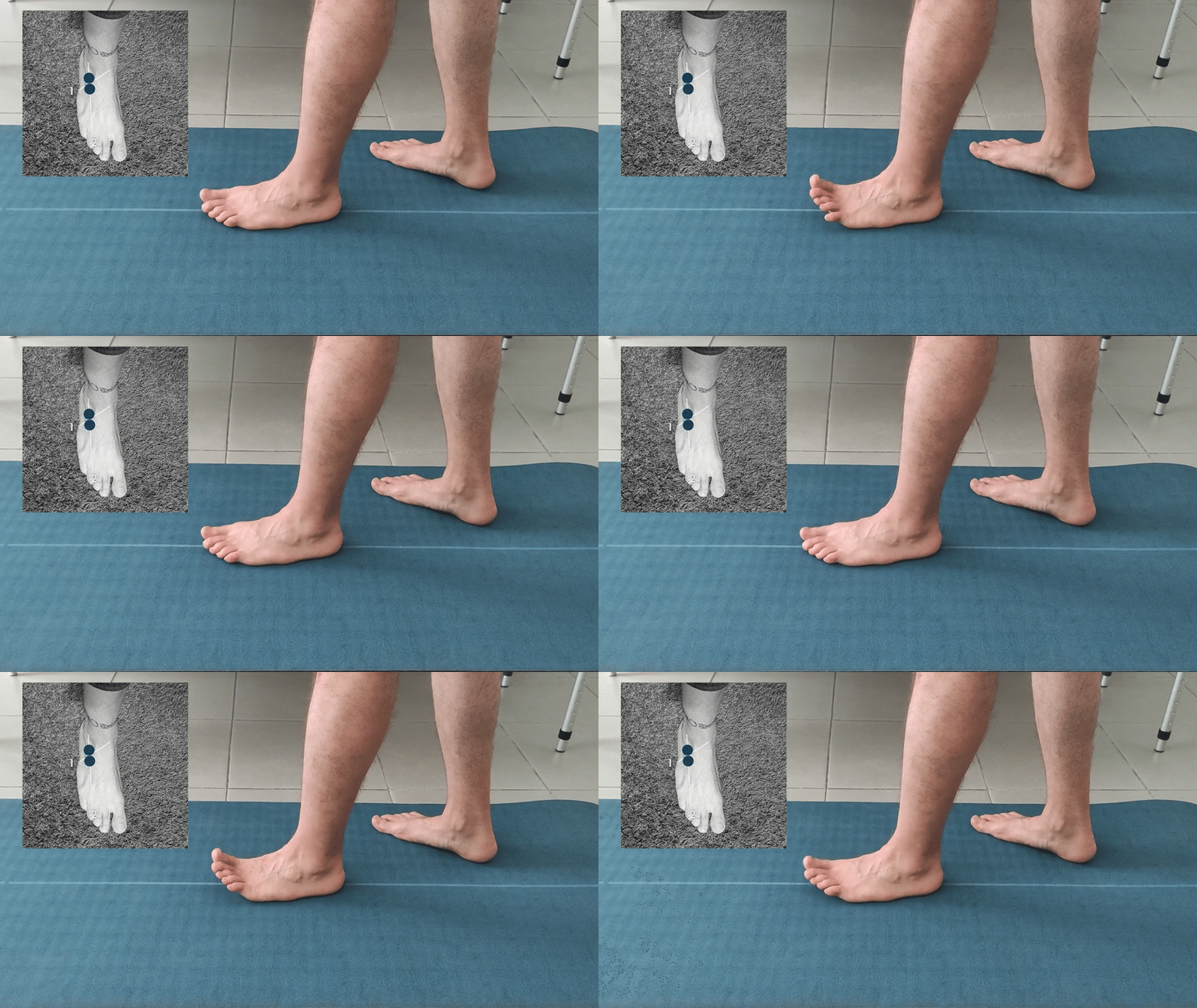
Function: extension of the proximal phalanges.
The extensor digitorum brevis acts as a toe extensor from the dorsum of the foot. Its function is essential for push-off during gait and for keeping the toes in a functional position during the stance phase.
When to assess it:
- – Dorsal foot pain.
- – Patients who do not place their toes on the ground, with flexed toes or claw toes.
- – Functional deficits of the first toe. In these cases, it makes sense to assess it in synergy with the flexor hallucis brevis to understand the balance between both muscles.
2. First dorsal interosseous
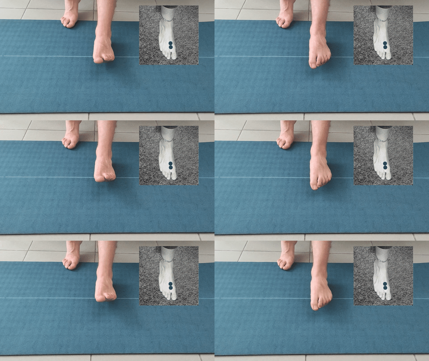
Function: flexion of the proximal phalanx and extension of the second and third phalanges of the second toe. Also involved in toe abduction.
When to assess it:
- – Patients with flexed and claw toes.
- – When there is limited toe separation, especially between the first and second toe.
The first dorsal interosseous is one of the least assessed muscles in standard clinical practice, but its deficit frequently appears in patients with forefoot dysfunction and alterations in fine digital movement control.
3. Abductor digiti minimi
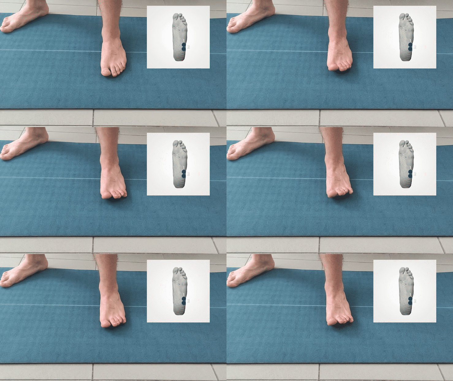
Function: abduction of the fifth toe.
The abductor digiti minimi stabilises the lateral border of the foot and contributes to lateral control during gait and single-leg stance.
When to assess it:
- – Deficit in toe separation, especially if the fifth toe is affected.
- – Patients with pain on the lateral border of the foot or lateral instability during gait.
4. Flexor digitorum brevis
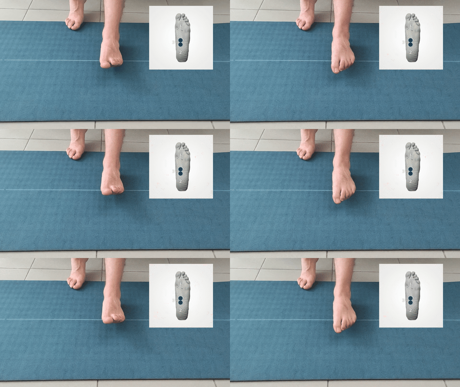
Function: flexion of the four lateral toes (second to fifth) and plantar flexion of the foot.
The flexor digitorum brevis is one of the most relevant muscles in plantar arch stabilisation. It works continuously during the stance phase and push-off, and its deficit is associated with some of the most common plantar pathologies seen in clinical practice.
When to assess it:
- – Pain during toe contact with the ground.
- – Plantar fasciopathies.
- – Flatfoot or cavus foot.
- – Calcaneal spur.
In all these cases, EMG allows you to verify whether the flexor digitorum brevis is generating sufficient activation to contribute to arch support or whether the load is falling excessively on the plantar fascia.
5. Flexor hallucis brevis
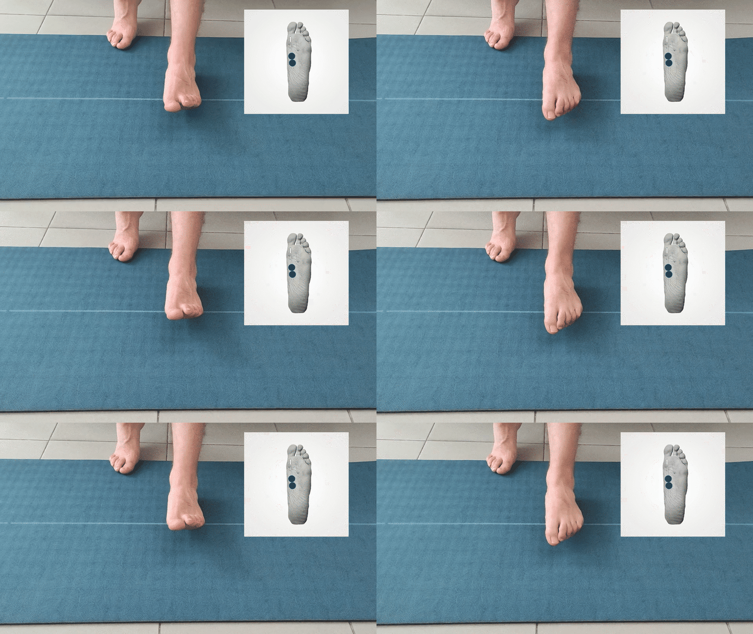
Function: flexion of the proximal phalanx of the first toe at the metatarsophalangeal joint.
The flexor hallucis brevis is key for push-off during walking and running. Its dysfunction directly affects step efficiency and can generate compensatory patterns that ascend through the kinetic chain.
When to assess it:
- – Functional deficits of the first toe: hallux rigidus, hallux valgus, metatarsophalangeal joint dysfunction.
- – Alterations in gait, running or functional toe loading.
Whenever you assess the extensor digitorum brevis, it also makes sense to include the flexor hallucis brevis to analyse the balance between both antagonists. A flexor deficit can explain first toe hyperextension; an extensor digitorum brevis deficit, sustained flexion.
Frequently asked questions
Is it difficult to place electrodes on the foot muscles? Foot musculature requires precision in electrode positioning, especially for smaller muscles such as the first dorsal interosseous or the abductor digiti minimi. The mDurance application includes specific placement guides for each muscle, orienting you on the exact electrode position.
Can I assess these muscles dynamically or only statically? You can assess them in both conditions. Static assessment allows you to isolate the function of each muscle. Dynamic assessment, during gait or functional exercises, shows how they behave under real loading conditions. Combining both perspectives gives the most complete picture of the deficit.
Is surface EMG valid for such small muscles? Yes, though with some nuance. In very small or deep muscles, crosstalk from adjacent muscles is greater. This is why precise electrode positioning and contextualised data interpretation are especially important when assessing foot musculature.
For which pathologies is this assessment most useful? Plantar fasciopathies, calcaneal spur, hallux valgus, flatfoot, cavus foot and gait alterations are the contexts where EMG assessment of the intrinsic foot musculature provides the most clinically actionable information.
Conclusion
The intrinsic musculature of the foot plays a determining role in arch stability, gait and joint health throughout the entire lower limb.
Yet it is one of the regions where objective assessment is most notably absent in standard clinical practice.
With mDurance you can measure the real activation of the extensor digitorum brevis, first dorsal interosseous, abductor digiti minimi, flexor digitorum brevis and flexor hallucis brevis.
You can compare sides, detect which muscle is failing, understand the synergies and design a treatment programme based on what is really happening, not on what you estimate should be happening.
Because in the foot, as in any other region, the difference between a good diagnosis and an incomplete one often lies in what you cannot see with the naked eye.