Rectus Femoris–Biceps Femoris Synergy
The balance between the rectus femoris and biceps femoris is one of the most important synergies in the biomechanics of both walking and running.
When these muscles work in harmony, the leg efficiently absorbs impact, controls the knee, and generates powerful propulsion.
But when this synergy is disrupted, the body enters a cycle of compensations, overloads, and inefficiencies that can lead to injuries such as patellar tendinopathy, hamstring strains, or patellofemoral pain.
That’s why being able to measure and analyze this synergy using surface electromyography (sEMG) is one of the most powerful tools you can incorporate into your clinical or sports rehabilitation practice.
👉 Want to learn how to apply EMG protocols to analyze running patterns and optimize your treatments?
Write to us here and discover how mDurance can help you measure, interpret, and retrain muscle activation with scientific precision.
Problems Associated with a Rectus Femoris > Biceps Femoris Activation Pattern
When the rectus femoris dominates—activating more than the biceps femoris—the movement pattern becomes stiff and inefficient.
This imbalance can appear at different phases of the gait or running cycle and has major implications for both injury prevention and performance.
Most common effects:
❌ Loss of eccentric knee control during stance → hamstrings fail to decelerate extension, increasing patellar tendon load.
❌ Quadriceps overload and risk of patellar tendinopathy → rectus femoris takes on too much work during impact absorption.
❌ Reduced shock absorption at initial contact → the leg becomes “stiffer,” transmitting more impact to the knee and hip.
❌ Poor hamstring coordination and disuse → biceps femoris loses influence, limiting knee control and hip extension.
❌ Lower propulsion efficiency → with poor hamstring contribution, the final push-off weakens, reducing speed and running economy.
Why You Should Evaluate This Synergy with EMG
It’s a cliché, but true: what isn’t measured can’t be corrected.
Visual observation or video analysis alone cannot reveal what’s happening at the neuromuscular level.
Surface electromyography (sEMG) allows you to measure actual muscle activation in real time, during movement.
This lets you identify whether your patient or athlete:
⚠️ Overuses the quadriceps and underactivates the hamstrings.
⚠️ Shows asymmetries between legs.
⚠️ Needs to adjust technique or retrain neuromuscular control before progressing in load or speed.
Step-by-Step Assessment Protocol
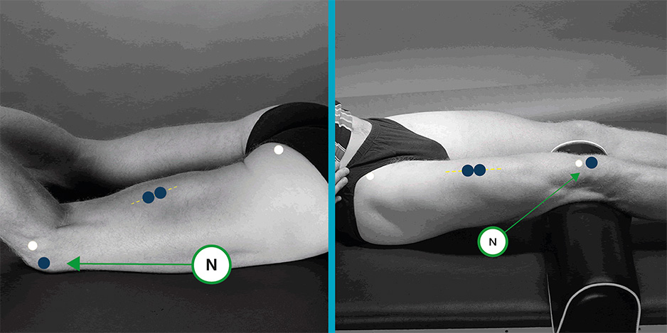
1. Electrode Placement
For accurate readings:
- Rectus femoris: place electrodes at the midpoint of the thigh, along the line between the anterior superior iliac spine and the upper patellar border.
Biceps femoris: place electrodes over the muscle belly, midway down the posterior thigh, aligned with the fiber direction.
2️. Evaluate Walking First
Have the patient walk at a natural pace. This provides a baseline to detect asymmetries.
Under normal conditions, quadriceps and hamstrings activation should alternate during opposite phases of the gait cycle:
- During stance phase, the rectus femoris controls knee flexion.
- During swing phase, the biceps femoris acts eccentrically to decelerate extension and prepare for the next contact.
3. Evaluate During Running
Once the baseline pattern is established, repeat the assessment during a light or moderate run.This phase highlights the most relevant imbalances, since increased velocity and load demand greater neuromuscular coordination.
Real EMG Patterns
✅ Normal Running Pattern
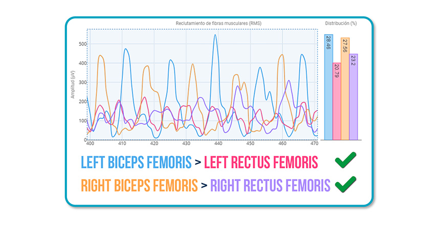
- Left biceps femoris > Left rectus femoris (V)
- Right biceps femoris > Right rectus femoris (V)
Clinical interpretation: This pattern shows good intermuscular coordination and effective eccentric knee control, significantly reducing injury risk.
❌ Altered Running Pattern
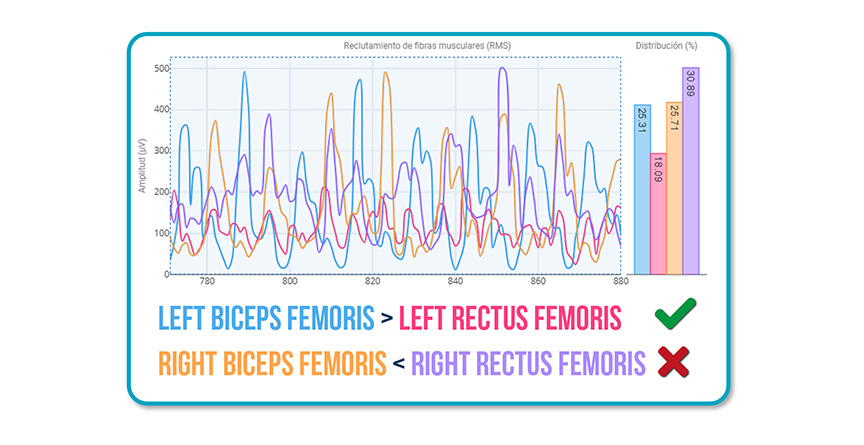
- Left biceps femoris > Left rectus femoris (V)
- Right rectus femoris > Right biceps femoris (X)
Here, the right rectus femoris is compensating for weak biceps femoris activation.
👉 Result: quadriceps overload, loss of eccentric control, and higher risk of tendon injury.
Clinical interpretation: This pattern is common after a hamstring injury or a period of inactivity.
Clinical Implications
The quadriceps-hamstring synergy affects not only sports performance but also recovery from knee, hip, or pelvic injuries.
Measuring this relationship with EMG enables you to:
✅ Detect activation deficits or post-injury compensations.
✅ Plan a return-to-running program with objective criteria.
✅ Design exercises and loads adapted to each patient’s true neuromuscular status.
See you in the next post 🙂