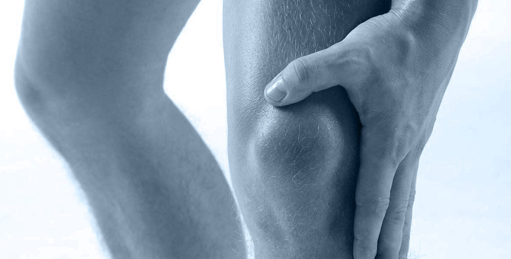
Lateral trunk rotation is a fundamental movement in human biomechanics, where the torso rotates to one side without excessive pelvic displacement.
This movement is crucial in daily activities such as turning to look behind, reaching for objects laterally, or performing tasks like driving. Additionally, it is essential in sports like golf, tennis, and baseball, where efficient rotational mobility is key to performance and injury prevention.
This movement primarily involves the external and internal obliques, spinal erectors, among others. Proper activation and synergy among these muscles ensure smooth and safe rotation, preventing compensations that could lead to overload in the lumbar or thoracic region.
Ineffective or uncoordinated muscle function in lateral trunk rotation can contribute to pain, stiffness, or postural imbalances. If you have patients experiencing mobility restrictions, weakness, or discomfort when rotating their trunk, the key question to ask is:
Which muscles are not functioning correctly to allow my patient to perform lateral rotation with optimal movement quality?
In this post, you will learn how to evaluate muscle synergies in lateral trunk rotation, identify imbalances, and tailor your treatment precisely to the muscles that need retraining.
By the way, if you want to enhance the accuracy of your assessments and optimize patient treatments, discover how mDurance’s EMG can help you.
When to Evaluate Lateral Trunk Rotation in a Patient
It is essential to assess lateral trunk rotation in your patients when they present any of the following symptoms or conditions:
- 1️⃣ Lumbar or thoracic pain: Any patient experiencing discomfort when rotating the torso, especially in the mid or lower back, should be evaluated. This may be related to muscular dysfunctions, joint blockages, or incorrect activation of stabilizing muscles.
- 2️⃣ Limited range of motion: If your patient struggles to rotate their trunk laterally to one side or feels stiffness while doing so, they may have mobility restrictions. This is common in cases of vertebral dysfunctions, muscle stiffness, or postural imbalances.
- 3️⃣ Previous trauma or surgery: Patients with a history of spinal injuries, abdominal surgery, or lumbar procedures require periodic evaluation of trunk rotation to monitor recovery and prevent compensations that could lead to new discomfort.
- 4️⃣ Muscle imbalances or postural deviations: Patients with altered postures, such as lumbar hyperlordosis, thoracic kyphosis, or pelvic asymmetries, may develop trunk rotation restrictions. Evaluating this function is key to improving mobility and preventing overloads.
- 5️⃣ Athletes relying on torso rotation: Athletes in sports like golf, tennis, baseball, or martial arts require precise evaluation of lateral rotation to prevent overuse injuries and ensure proper core muscle balance.
If you identify any of these signs in your patients, conducting a detailed assessment of lateral trunk rotation will help you detect limitations and design a more effective treatment plan.
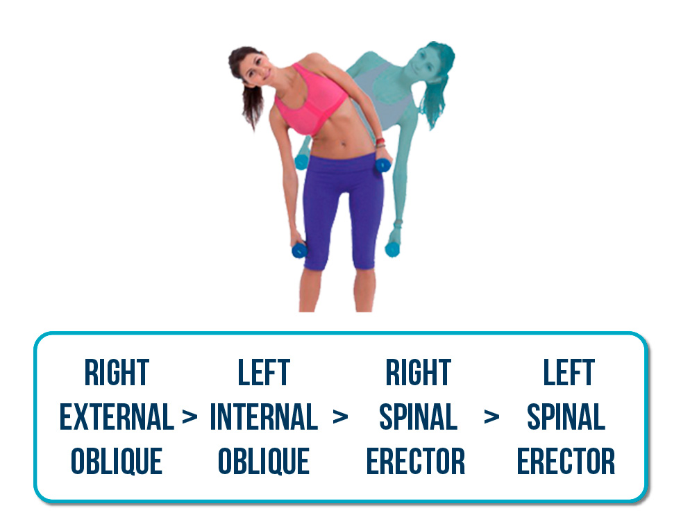
Muscles Most Active in Lateral Trunk Bending (to the Right)
- 1. Right external oblique: Pulls the ribcage toward the pelvis on the right side, facilitating lateral bending to that side.
- 2. Left internal oblique: Works to stabilize the trunk and assist in abdominal cavity compression.
- 3. Right spinal erector: Plays an essential role in maintaining an upright and stabilized spine during movement, preventing unwanted flexion or rotation.
- 4. Left spinal erector: Works similarly but to a lesser extent.
Expected Synergy in Right-Side Lateral Trunk Bending:
How to Assess This Synergy in Lateral Trunk Rotation
Example: Patient with Lumbar Pain
When analyzing muscle activation in lateral trunk rotation, it is crucial to observe the synergy among the involved muscles. In this case, the patient exhibits the following pattern:
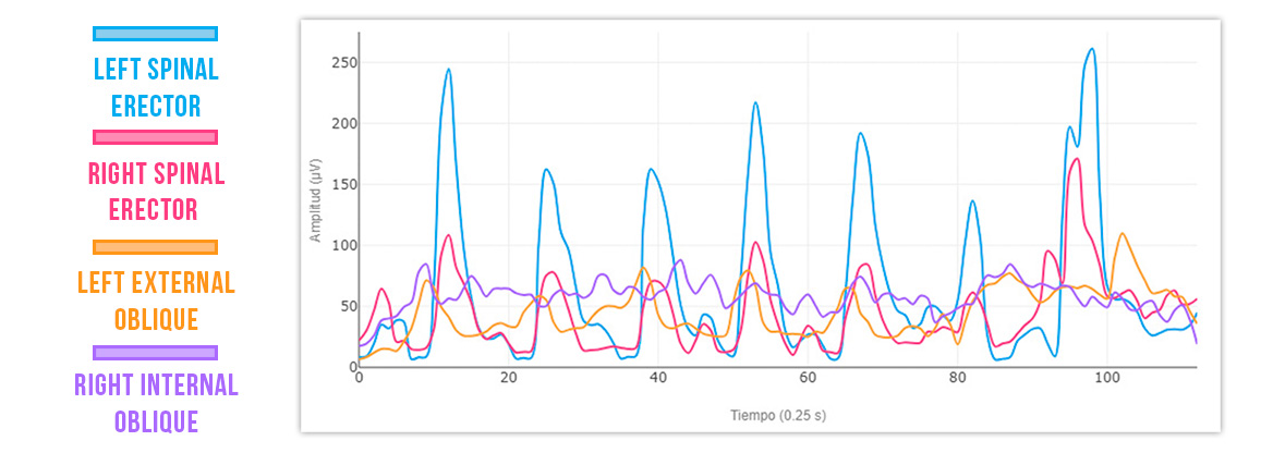
Left spinal erector > Right spinal erector > Left internal oblique > Right external oblique ❌
🔹 Excessive left spinal erector coactivation → Indicates overcompensation, which can cause overload and lumbar discomfort.
🔹 Deficient activation of the left internal oblique and right external oblique → Suggests inadequate abdominal muscle participation in rotation, reducing movement efficiency.
This dysfunctional pattern can lead to chronic lumbar pain, core instability, and compensations in movement mechanics. Identifying these imbalances with surface electromyography (EMG) allows for more precise and effective treatment strategies.
Example: Normal Activation Pattern
A proper muscle activation pattern is crucial for efficient and pain-free lateral trunk rotation. In a pain-free individual, muscle synergy follows this order:
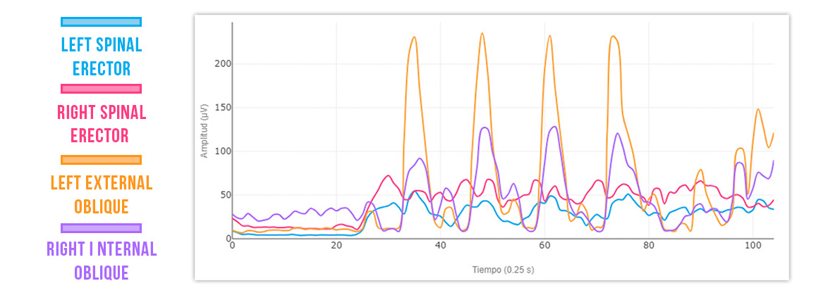
Right external oblique > Left internal oblique > Right spinal erector > Left spinal erector
This balance enables a stable and effective rotation, where the obliques lead the movement, and the spinal erectors provide stabilization without causing overload.
🚨 Comparison Between a Normal Pattern and a Patient with Lumbar Pain:
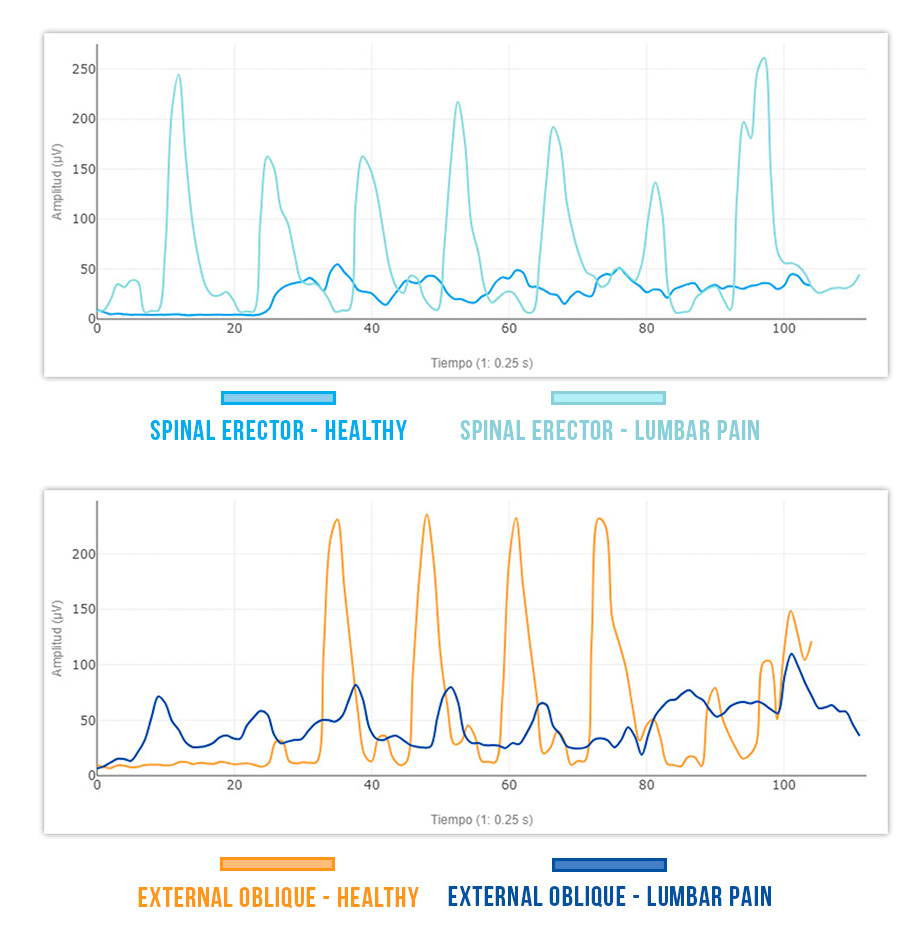
🔹 The left spinal erector in a patient with lumbar pain activates 3 times less than in a healthy person ❌ → This low activation can compromise stability and cause compensations in other muscles.
🔹 The healthy external oblique activates 3.5 times more than the external oblique of a patient with lumbar pain ❌ → A deficit in oblique activation reduces movement efficiency and increases lumbar stress
Conclusion
Understanding muscle activation during lateral trunk bending is key to evaluating and optimizing this movement. A detailed analysis helps identify imbalances and compensations, aiding in the prevention and treatment of dysfunctions that may lead to pain or functional limitations.
In a proper activation pattern, the external and internal obliques should act as primary agonists, leading the movement with predominant activation. If other muscles, such as the spinal erectors, take on an excessive role, compensations may occur, reducing movement efficiency and increasing overload risk.
Thanks to tools like surface electromyography (EMG), it is possible to objectively measure muscle activation and correct dysfunctional patterns.
If you want to enhance the accuracy of your assessments and optimize your patients’ treatments, discover how mDurance’s EMG can help you.
See you in the next post 🙂

